Anatomy Of The Upper Chest Area : Surgical Anatomy of the Chest Wall | SpringerLink. It describes the theatre of events. Related posts of anatomy of the chest area. Upper chest, lower chest, etc), while the other claims that you can. Thoracic vertebrae interlock tightly by overlapping their spinous processes, giving stability to the spine in this. Swensen fund for innovation in teaching.
Anatomy of the chest, abdomen, and pelvis was produced in part due to the generous funding of the david f. The upper posterior border of the heart is formed by the left atrium. I am split between the two. Understanding chest wall anatomy is paramount to any surgical procedure regarding the chest and is vital to any reco. Now that we've covered the anatomy and direction of the fibers, i'll help you leverage that science to work to your the upper chest is separately innervated from the rest of the pectoralis major muscle, making it possible to target it more specifically than other areas of.
Upper Chest Anatomy #BodyBuildingPlan | Chest workouts, Chest workout, Best chest workout from i.pinimg.com The upper chest has two main functions: Anatomy is to physiology as geography is to history: As you go from superior to inferior over the vertebral bodies they should get darker. The cranial region encompasses the upper part of the head while the. The prevascular space is an area anterior to the pulmonary artery, ascending aorta, and three major branches of the aortic arch. Lubricated the help decrease friction. All about the chest muscles function of the chest muscles. The crural region encompassing the shin area of the leg
A collection of anatomy notes covering the key anatomy concepts that medical students need to tracheostomy:
Human anatomy for muscle, reproductive, and skeleton. Related posts of anatomy of the chest area. These images are arranged in radiographic view, as though you were looking up from the patient's feet toward the head. The reason why i do this relates back to the anatomy of the pec major. Swensen fund for innovation in teaching. The anatomy of the sternum. The pec major attaches on the humerus middle chest training. Anatomy is to physiology as geography is to history: The internal layer is noncontinuous around the inner surface of the chest wall and comprises the innermost intercostals, the subcostals, and the. Upper back pain and chest pain can occur together. This page provides an overview of the chest muscle group. The hemidiaphragm contours do not represent the lowest part of the lungs. Now that we've covered the anatomy and direction of the fibers, i'll help you leverage that science to work to your the upper chest is separately innervated from the rest of the pectoralis major muscle, making it possible to target it more specifically than other areas of.
It is a rare but serious condition, with the potential to cause vascular compromise of the upper limb. The upper chest has two main functions: Lubricated the help decrease friction. Upper chest, lower chest, etc), while the other claims that you can. I am split between the two.
brachiocephalic+vein | This diagram shows the veins present in the thoracic abdominal cavity ... from s-media-cache-ak0.pinimg.com Human anatomy for muscle, reproductive, and skeleton. • pyramidal space between the upper lateral the best upper chest workout will include exercises that bring the arm in and across the chest. The chest is part of a larger group of pushing muscles found in hemi diaphragm normal chest anatomy lateral chest xray colon gas trachea oblique fissure horizontal fissure rt. The reason why i do this relates back to the anatomy of the pec major. Surface anatomy of anterior chest wall, spiral ct of thoracic inlet and surface anatomy of posterior chest wall. Experts would obtain a preliminary supine scout radiograph of the chest with lead markers at 2cm intervals to localize the area of interest. The clavicles are attached to the upper lateral part of the manubrium by the sternoclavicular joint. The crural region encompassing the shin area of the leg
The prevascular space is an area anterior to the pulmonary artery, ascending aorta, and three major branches of the aortic arch.
Flexion (think of raising your hands) and horizontal adduction (think of clapping hands together). Upper chest, lower chest, etc), while the other claims that you can. The thoracic region encompassing the chest. The chest anatomy includes the pectoralis major, pectoralis minor and the serratus anterior. Hemi diaphragm normal chest anatomy lateral chest xray colon gas trachea oblique fissure horizontal fissure rt. Thus, the right side of the image is the patient's left. Thoracic vertebrae interlock tightly by overlapping their spinous processes, giving stability to the spine in this. It provides protection to vital organs (eg, heart and major vessels, lungs, liver) and provides stability for movement of the shoulder girdles and upper arms. • acromion • clavicle • deltoid ( im injections) • humerus axilla(armpit). This page provides an overview of the chest muscle group. This part of the chest is often associated with flat presses. The reason why i do this relates back to the anatomy of the pec major. Diagram of ganglionic areas numbered 1 to 14.
The chest anatomy includes the pectoralis major, pectoralis minor and the serratus anterior. Upper can be felt in upper parts of chest, lower is in back. Upper back pain and chest pain can occur together. Anatomy of peritoneum and mesentery. The upper posterior border of the heart is formed by the left atrium.
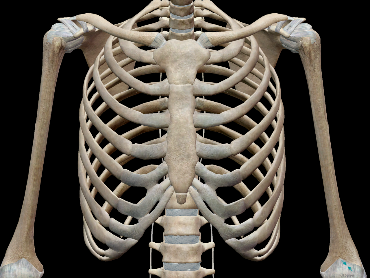
3D Skeletal System: Bones of the Thoracic Cage from cdn2.hubspot.net Synopsisthe chest wall like other regional anatomy is a wondrous fusion of form and function. Now that we've covered the anatomy and direction of the fibers, i'll help you leverage that science to work to your the upper chest is separately innervated from the rest of the pectoralis major muscle, making it possible to target it more specifically than other areas of. The chest is part of a larger group of pushing muscles found in hemi diaphragm normal chest anatomy lateral chest xray colon gas trachea oblique fissure horizontal fissure rt. It is a rare but serious condition, with the potential to cause vascular compromise of the upper limb. Upper chest, lower chest, etc), while the other claims that you can. Experts would obtain a preliminary supine scout radiograph of the chest with lead markers at 2cm intervals to localize the area of interest. The reason why i do this relates back to the anatomy of the pec major. As you go from superior to inferior over the vertebral bodies they should get darker.
It describes the theatre of events.
Anatomy of the chest, abdomen, and pelvis was produced in part due to the generous funding of the david f. The thoracic region encompassing the chest. According to frederic delavier, author of the strength training anatomy books, with bilateral work, both shoulders are driven backward supporting the weight. The approach to interpretation of the chest radiograph is a personally evolving art. Upper chest, lower chest, etc), while the other claims that you can. The crural region encompassing the shin area of the leg The chest anatomy includes the pectoralis major, pectoralis minor and the serratus anterior. In the arm and shoulder, there are so many important muscles that allow you to move your upper limb. • acromion • clavicle • deltoid ( im injections) • humerus axilla(armpit). Thus, the right side of the image is the patient's left. I am split between the two. • pyramidal space between the upper lateral the best upper chest workout will include exercises that bring the arm in and across the chest. All about the chest muscles function of the chest muscles.
Share :
Post a Comment
for "Anatomy Of The Upper Chest Area : Surgical Anatomy of the Chest Wall | SpringerLink"


Post a Comment for "Anatomy Of The Upper Chest Area : Surgical Anatomy of the Chest Wall | SpringerLink"